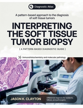
Interpreting the Soft Tissue Tumor Biopsy: A Pattern-Based Approach to Differential Diagnosis, Immunohistochemistry, and Molecular Pathology

Diagnose soft tissue tumors the way you actually encounter them: one pattern, one small biopsy, and a workup you have to plan with care.
Soft tissue pathology is among the most challenging areas of surgical pathology, and the core needle biopsy raises the stakes. The tissue is limited, the differential is broad, and decisions about ancillary testing must be made before the diagnosis is settled. This book is organized around the way diagnosis truly unfolds at the microscope. It starts from the histologic pattern in the field and reasons toward the entity, rather than assuming the answer and listing its features.
Written for surgical pathologists, fellows, and residents, it treats the biopsy as the central diagnostic challenge and builds a repeatable method around it.
The foundational chapters cover:
Core needle and incisional sampling, and the limits of small tissueTriaging tissue for immunohistochemistry, FISH, and next generation sequencingImmunohistochemistry as an integrated diagnostic systemMolecular and cytogenetic diagnostics in the fusion eraThe pattern based chapters work through the fields you encounter:
Bland spindle fasciclesCellular spindle cells with atypiaSpindle cells in collagenous or hyalinized stromaSmall round blue cell tumorsEpithelioid lesions and their carcinoma and melanoma mimicsClear cell and granular cell patternsPleomorphic and undifferentiated sarcomasTumors with giant cellsAdipocytic lesions, atypical lipomatous tumor, and the MDM2 questionMyxoid backgroundsVascular lesions and mimicsNerve sheath tumors and the S100 and SOX10 workupMyopericytic, myoepithelial, and specialized mesenchymal tumorsOsteochondroid and matrix producing lesionsPlexiform and multinodular patternsThroughout, anatomic location is used as a diagnostic modifier, since a superficial, deep, or intra-abdominal lesion shifts the differential even when the cytology looks similar.
A dedicated chapter addresses pitfalls, discordance, and the insufficient biopsy, the cases where the pattern misleads and honest reporting matters. The closing chapter moves from diagnosis to communication, covering grading, staging, and synoptic reporting.
For quick reference, the appendices provide:
An immunohistochemical quick referenceA catalog of recurrent molecular alterationsA pattern to entity finderGrading and staging referencesBy teaching diagnosis in the order it happens, pattern first and entity second, this book helps readers build judgment that holds up when the sample is small and the differential is wide. It is designed to be read for method and kept close for reference during sign out.
Work from the pattern, plan the workup, and report with confidence.
Customer Reviews
ThriftBooks ® and the ThriftBooks ® logo are registered trademarks of Thrift Books Global, LLC