


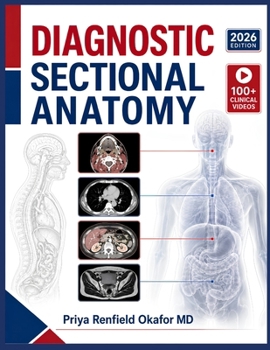
Diagnostic Sectional Anatomy: Image Quality Assessment, Structure Differentiation, Slice Positioning Accuracy, and Evidence-Based Interpretation for I

Struggling to tell true pathology from a scanner artifact at 3 AM? Most imaging residents can name a structure on a textbook diagram but freeze the moment a real cross-sectional study shows noise, beam hardening, or a partial-volume shadow that mimics disease.
Every year, promising residents fall behind not because they lack anatomic knowledge, but because no one ever taught them how image quality, slice positioning, and reformatting technique change what a structure actually looks like on the screen in front of them.
Diagnostic Sectional Anatomy closes that gap. Written by a practicing radiologist for the 2026 academic year, this reference walks through the physics of CT and MRI image quality, systematic search patterns, and region-by-region sectional anatomy from the brain to the ankle, always tying structure back to how it is actually acquired, reformatted, and reported.
25 chapters covering imaging physics, structured search technique, and every major anatomic region in sectional detailClinical decision points, differential-diagnosis mini-tables, and quick-reference sidebars built for real interpretive practiceOver 45 detailed clinical illustrations showing exact views, labeled structures, and color-coded anatomyA companion video course of more than 50 curated lectures, accessible instantly through the QR code printed insideEvidence-based coverage of structured reporting, discrepancy reduction, and advanced 3D post-processing toolsWhether preparing for boards, starting a radiology rotation, or building the systematic search habits that prevent missed findings, this 2026 edition gives residents a single, coherent framework for reading sectional imaging with confidence.
Stop guessing at the screen. Start reading it like a radiologist.
Customer Reviews
ThriftBooks ® and the ThriftBooks ® logo are registered trademarks of Thrift Books Global, LLC