


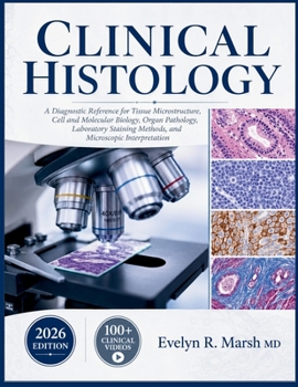
Clinical Histology: A Diagnostic Reference for Tissue Microstructure, Cell and Molecular Biology, Organ Pathology, Laboratory Staining Methods, and Mi

Struggling to connect a stained slide to the diagnosis it points toward? Most histology references stop at labeling structures. They leave the harder skill, reading tissue the way a working pathologist does, entirely unaddressed.
This 2026 edition closes that gap. Across 25 chapters, it walks from foundational microscopy and cell biology through every major organ system, pairing each tissue's normal architecture with the diagnostic patterns clinicians actually encounter on biopsy and autopsy.
Clinical pearls in every subchapter, distilled from real diagnostic reasoningCase vignettes with worked differential-diagnosis logicOver 50 detailed clinical illustrations spanning normal and pathologic microstructureStaining protocols, immunohistochemistry, and modern molecular pathology techniquesQuick-reference tables and decision points built for fast recall under exam or clinical pressureIf you have ever stared down a microscope unsure which structure you were even looking at, this book gives you the framework to read tissue with confidence.
A QR code links every reader to a companion video course of over 100 curated lectures, reinforcing each chapter with real recorded teaching alongside the illustrations inside these pages.
Clinical Histology was built for medical students, pathology residents, laboratory scientists, and any clinician who wants their microscope findings to translate directly into diagnostic confidence.
Customer Reviews
ThriftBooks ® and the ThriftBooks ® logo are registered trademarks of Thrift Books Global, LLC