


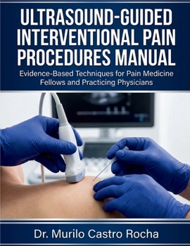
Ultrasound-Guided Interventional Pain Procedures Manual: Evidence-Based Techniques for Pain Medicine Fellows and Practicing Physicians

This procedural manual provides instruction for 39 ultrasound-guided interventional pain techniques performed in clinical practice. The textbook addresses diagnostic and therapeutic nerve blocks, joint injections, and spinal procedures across six anatomical regions.
Content Organization
The manual contains 39 chapters addressing foundational principles and procedural techniques:
Chapters 1-6: Foundational Principles cover ultrasound physics and machine optimization, sonoanatomy fundamentals, sterile technique and infection prevention, local anesthetic pharmacology and systemic toxicity, patient selection and informed consent, and complications including recognition, prevention, and management.
Chapters 7-11: Head and Neck Procedures include greater occipital nerve block, third occipital nerve block, cervical medial branch blocks, stellate ganglion block, and superficial cervical plexus block.
Chapters 12-18: Upper Extremity Procedures cover suprascapular nerve block, glenohumeral joint injection, acromioclavicular joint injection, subacromial-subdeltoid bursa injection, lateral epicondyle injection (tennis elbow), medial epicondyle injection (golfer's elbow), and radial nerve block at the elbow.
Chapters 19-23: Thoracic and Truncal Procedures address intercostal nerve blocks, thoracic paravertebral blocks, thoracic medial branch blocks, transversus abdominis plane (TAP) block, and ilioinguinal and iliohypogastric nerve blocks.
Chapters 24-30: Lumbar Spine and Pelvic Procedures include lumbar medial branch blocks, lumbar facet joint injections, sacroiliac joint injection, caudal epidural injection, interlaminar lumbar epidural injection, piriformis muscle injection, and ganglion impar block.
Chapters 31-39: Lower Extremity Procedures cover lateral femoral cutaneous nerve block, femoral nerve block, sciatic nerve block (subgluteal approach), hip joint injection (anterior approach), trochanteric bursa injection, genicular nerve blocks, knee joint injection, common peroneal nerve block, and tibial nerve block at the ankle.
Chapter Structure
Each procedural chapter follows a standardized format:
Clinical indications with diagnostic criteriaAnatomical overview with cross-sectional relationshipsSonoanatomic landmarks and tissue identificationPatient positioning requirementsUltrasound probe selection and placementTarget structure identification on ultrasoundNeedle approach techniques (in-plane and out-of-plane)Injection volumes and medication selectionConfirmation methods for accurate placementComplications with recognition and managementEvidence-based outcomes from current literaturePost-procedure care protocolsClinical Feature Boxes
Each chapter includes structured educational elements:
Clinical Pearls provide ultrasound optimization strategies, needle visualization techniques, anatomical identification tips, and procedural refinements based on clinical experience.
Equipment Checklists detail ultrasound equipment specifications, sterile supplies, needle gauge and length requirements, syringe sizes, medications with dosing, monitoring equipment, and emergency supplies.
Patient Safety Alerts identify absolute and relative contraindications, critical neurovascular structures to avoid with specific avoidance strategies, local anesthetic dosing limits, high-risk patient populations, and preprocedural verification requirements.
Evidence-Based Medicine Boxes
Treatment Algorithms
Differential Diagnosis Tables
Diagnostic Criteria Tables
Emergency Management Protocols
Customer Reviews
ThriftBooks ® and the ThriftBooks ® logo are registered trademarks of Thrift Books Global, LLC