Cranial Nerves for Medical Students: with clinical correlations

Cranial Nerves for Medical Students: Anatomy with Clinical Correlations
by Dr. Michael M. Nikoletseas
(Oxford University Library)
Cranial Nerves for Medical Students: Anatomy with Clinical Correlations by Dr. Michael M. Nikoletseas is a specialized educational resource designed to enhance understanding of the cranial nerves through a three-dimensional and clinically integrated approach. The book is particularly valuable for medical education because it bridges anatomical learning with practical clinical applications. It is accessible through major academic and online platforms.
Book Content and Structure
The book focuses on teaching the cranial nerves in three-dimensional anatomical space, a method that provides knowledge essential for practicing physicians. It presents the pathway of each nerve in fine detail, taking the reader on a virtual walk along its entire course from origin to termination. This approach contrasts with traditional instructional methods that rely primarily on lists, tables, and mnemonics, which are considered less effective for clinical practice.
An important feature of the book is its integration with neuroanatomy, which is traditionally taught in the second semester of the first year of medical school in the United States. This organization supports a smoother transition between gross anatomy and neuroanatomy and addresses gaps commonly found in standard curricula. The book also includes detailed tracings of cranial nerves within the cranium, making it particularly useful for neurosurgeons and radiologists.
Clinical Correlations
The text emphasizes clinical correlations throughout. All twelve cranial nerves are presented with clinical applications, linking anatomical structure to functional deficits and pathological conditions. This emphasis supports the practical use of anatomical knowledge in diagnosis and treatment.
Cranial Nerves for Medical Students: Anatomy with Clinical Correlations is a comprehensive and clinically oriented guide for students seeking a deeper understanding of both the anatomical and clinical aspects of the cranial nerves. Its integration of detailed anatomy with practical applications, supported by high-quality illustrations from Gray's Anatomy, makes it especially useful in dissection laboratories and neurosurgical contexts.
Facts about This Book
The book covers a specific part of gross anatomy: the cranial nerves of the head and neck.
The author has taught this material as head of the Head and Neck unit.
The author has conducted research in neuroanatomy and neurophysiology.
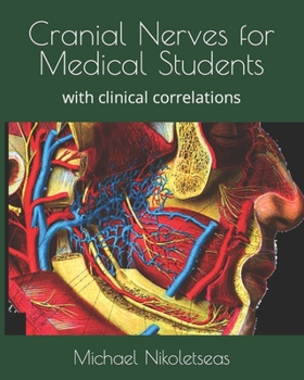
The illustrations are drawn from Gray's Anatomy, emphasizing classical anatomical accuracy rather than decorative style.
The cranial nerves and the cranium are presented in minute anatomical detail.
Each nerve is followed centimeter by centimeter from origin to termination.
The book contains original color illustrations depicting the complex nerve pathways in detail.
The text can be used in the dissection laboratory.
Clinical correlations are presented for all twelve cranial nerves.
The type of knowledge developed is essential for diagnosis and treatment of difficult cases.
The detailed three-dimensional anatomical understanding provided is particularly necessary for neurosurgeons.
Background and Author Information
Dr. Michael M. Nikoletseas is the author of the book and has a background in medicine with extensive experience teaching medical anatomy in U.S. medical schools, including the University of Wisconsin-Madison. He has taught the Head and Neck unit and has conducted research in neuroanatomy and neurophysiology.
Customer Reviews
ThriftBooks ® and the ThriftBooks ® logo are registered trademarks of Thrift Books Global, LLC