


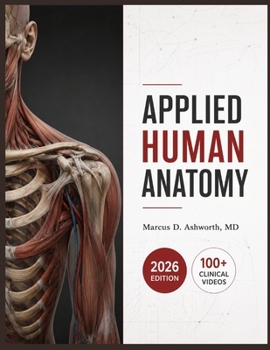
Applied Human Anatomy: A Clinical Illustrated Reference for Regional Dissection, Musculoskeletal Architecture, Neurovascular Pathways, Surgical Surfac

You memorized the diagrams. Then a real patient walked in and the textbook version didn't match what you were seeing.
Cadaver photos rarely resemble the body on the table or the scan on the monitor. Students and working clinicians alike lose hours reconciling color-coded diagrams against the gray, layered reality of dissection, ultrasound, and the operating field.
This 2026 edition closes that gap. Twenty-five chapters move region by region, from surface landmarks a clinician palpates through the deep neurovascular planes a surgeon must name before opening a cavity, anchored throughout to imaging findings and bedside correlations instead of description for its own sake.
Twenty-five clinically anchored chapters covering osteology, myology, the vascular and lymphatic systems, the brain and cranial nerves, thoracic, abdominal, and pelvic viscera, the reproductive organs, both limbs, and surgical surface anatomyOne custom clinical illustration per chapter with exact anatomical labelingClinical Decision Point boxes, differential-diagnosis mini-tables, and quick-reference sidebars throughoutCase vignettes tying structure to real diagnostic reasoningA free QR-linked video course of over fifty lecture videos organized to mirror the book's chaptersEvery structure is framed the way it actually appears in practice: as a landmark to palpate, a plane to dissect, or a shadow to interpret on a scan.
Scan the code inside, work through the companion videos alongside the text, and stop translating between the atlas and the patient.
Customer Reviews
ThriftBooks ® and the ThriftBooks ® logo are registered trademarks of Thrift Books Global, LLC